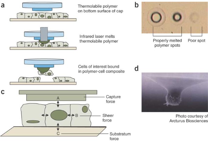
Laser capture microdissection lcm offers a rapid and precise method of isolating and removing specified cells from complex tissues for subsequent analysis of their rna dna or protein content thereby allowing assessment of the role of the cell type in the normal physiologic or disease process being studied.
Laser capture microdissection nature protocols.
This protocol enables amplification of the total transcript of a single prokaryotic cell for in depth analysis.
The use of laser capture microdissection lcm and quantitative polymerase chain reaction to define thyroid hormone receptor expression in human term placenta.
As opposed to xmd standard laser capture microdissection lcm is based on visual identification of target cells through a microscope 1 2.
Laser capture microdissection is used to isolate single cells and amplified cdna.
Methods and protocols third edition seeks to aid researchers generally and pathologists in particular in moving their studies forward with these vital techniques.
Laser capture microdissection lcm allows the isolation of specific cellpopulations from complex tissues that can be then used for gene expressionstudies.
Placenta 25 758 762 2004.
However there are no reproducible protocols to study rna in the brainand particularly in the substantia nigra.
Springer nature is developing a new tool to find and evaluate protocols.
2011 laser capture microdissection.
A histological section is stained and then a.
Laser capture microdissection lcm is a method to procure subpopulations of tissue cells under direct microscopic visualization.
Laser capture microscopy lcm coupled with global transcriptome profiling could enable precise analyses of cell populations without the need for tissue dissociation but has so far required.
Methods in molecular biology methods and protocols vol 755.
Cite this protocol as.
Decarlo k emley a dadzie o e mahalingam m.


